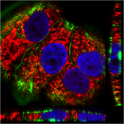
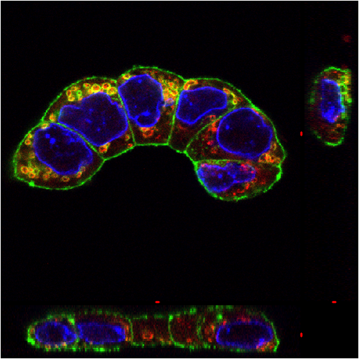
Comparison before (first image) and after (second image) exposure of anticancer agent to T47D breast cancer cell. Blue: nucleus, green: actin, red: mitochondria Specimen courtesy of Dr. Mitsuhiro Kudo, Department of Pathology, Nippon Medical School
코리아인스텍(주)|대표자 : 장삼섭 | 사업자등록번호 : 314-81-73532 | 개인정보관리책임자 : 조일현(8220282@naver.com)
주소 : 대전광역시 유성구 유성대로 828번길 52, 402호(장대동, 초산빌딩) | 대표번호 : 042)822-0282|팩스 : 042)823-0666
COPYRIGHT 2013 BY 코리아인스텍(주) ALL RIGHTS RESERVED


 3 color fluorescence
;)
;)


